No-Wash Homogeneous Assay
SPARCL® (Spatial Proximity Analyte Reagent Capture Luminescence) technology is a proximity dependent, non-separation, chemiluminescent detection method. In a SPARCL assay, a chemiluminescent substrate (acridan) is brought into the proximity of an oxidative enzyme (horseradish peroxidase, HRP) through the specific antigen/antibody interaction (Figure 1). A flash of light proportional to the quantity of analyte present in the sample is generated upon addition of a trigger solution containing H2O2 and para-hydroxycinnamic acid (pHCA). There is no need to remove excess reactants. This assay technology, applicable to both sandwich and competitive assays, has been implemented in formats with and without a solid phase. In the format with a solid phase, both the acridan compound and a specific capture antibody are coupled to solid phases such as micro particles or microtiter plates. Whereas when the solid phase is omitted the capture antibody is directly labeled with the acridan compound. Furthermore, to enhance signal to noise ratio, a background reducing agent can be added to minimize the background signal from unbound reactants.
Figure 1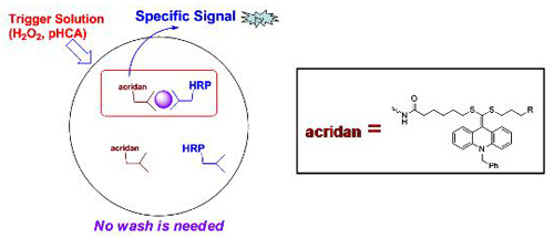
Plate-based SPARCL Assay*
To evaluate the SPARCL technology in a microtiter plate format an assay was performed with a white microtiter plate pre-coated with sheep anti-mouse IgG and acridan-labeled BSA. To the plate were added mouse IgG standard solution and goat anti-mouse IgG F(ab1)2-HRP. After incubation (1 hr), the plate was transferred to a Labsystem Model 391 Luminoskan** plate luminometer. Without removing the excess reagents, luminescence was generated by injecting a trigger solution and measured the light output (integrated for 5 seconds).
The standard curve from the mouse IgG assay are shown in figure 2.
Figure 2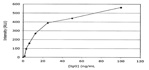
Microparticle-based SPARCL TSH Assay*
To evaluate the SPARCL technology in a microparticle based format, capture particles were prepared by covalently coupling mouse anti-TSH MAb and the acridan compound to Dynal M-270 amino-functionalized particles. Capture particles, HRP-conjugated monoclonal anti-TSH MAb and a TSH calibrator were combined and incubated for 1 hr in a 96-well plate. Chemiluminescence was generated by injection of trigger solution and integrated for 5 seconds on a Labsystem Model 391 Luminoskan** plate luminometer.
The standard curve from the microparticle-based SPARCL TSH assay is shown in figure 3.
Figure 3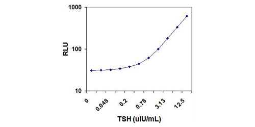
Solution-Phase SPARCL
To evaluate the SPARCL technology in the absence of a solid phase, specific antibody was directly labeled with acridan. TNF-α, IL-8, and PSA were analytes chosen to evaluate a sandwich assay format and cAMP was the analyte chosen to evaluate competitive assay format.
Solution-Phase SPARCL TNF-a Assay*
To evaluate SPARCL TNF-a assay in a solution phase format two complementary anti-TNF-a MAb antibodies were labeled, one with the acridan, and the other with biotin. The assay mixture, in a 96-well microtiter plate, contained 15µL acridan-labeled mouse anti-human TNF-a, 15µL biotinylated goat anti-human TNF-a, 30µL TNF-a standard solution and 15µL streptavidin-HRP. The mixture was incubated for 1 hour at room temperature then 10µL of ascorbic acid was added followed by the addition of 100µL trigger solution. Signal was read and integrated for 2 seconds post triggering on a Labsystem Model 391 Luminoskan** plate luminometer.
Figure 4 shows a typical standard curve for the TNF-a assay.
Figure 4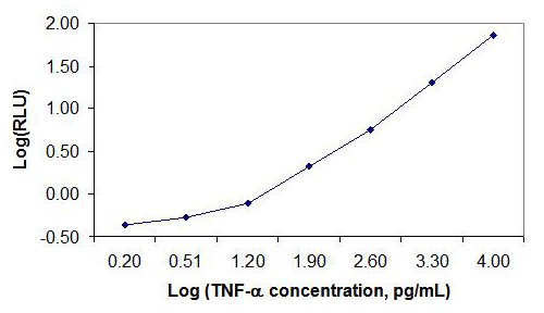
Solution-Phase SPARCL IL-8 Assay*
To evaluate SPARCL IL-8 assay in a solution phase format two complementary anti-IL-8 MAb antibodies were labeled, one with the acridan, and the other with HRP. No subsequent purification of unbound acridan was performed. The assay mixture, in a 96-well microtiter plate, contained 20µL acridan-labeled mouse anti-human IL-8 MAb, 20µL complementary HRP-anti-human IL-8 MAb and 30µL IL-8 standard solution. The mixture was incubated for 60 minutes at room temperature. A solution of ascorbic acid (10µL) was added followed by injection of trigger solution (100µL). Signal was read and integrated for 2 seconds post triggering on a Labsystem Model 391 Luminoskan** plate luminometer.
A typical standard curve for the IL-8 assay is shown in figure 5.
Figure 5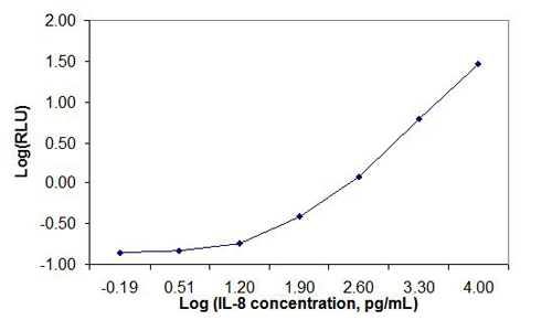
Solution-Phase SPARCL cAMP Assay*
The competitive cAMP assay, utilizing an acridan-labeled anti-cAMP MAb and HRP-cAMP as the competing binding molecule, was performed in 384-well plate format using Beckman Coulter BioRAPTR FRD** and Biomek-2000** to dispense the reagents. The reagents and assay conditions are as follows: 6µL acridan-labeled anti-cAMP MAb, 6µL HRP-cAMP and 4µL cAMP standard; 45 min incubation; 2µL ascorbic acid solution; 14µL trigger solution and 1-second signal integration on Molecular Devices SpectraMax-L**.
The cAMP competitive assay with a 45-minute incubation time achieved a calculated (2 standard deviations from background) analytical sensitivity of 0.274 nM and two orders of magnitude in dynamic range (Figure 6). The assay yielded an IC50 of 1.05nM. A 5-parameter logistics fit was done with Molecular Devices SoftMax Pro 5.4**. The assay exhibited a Z’ of 0.73.
Figure 6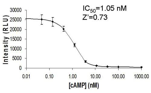
Solution-Phase SPARCL PSA Assay*
Anti-PSA MAb was labeled with the acridan compound. No subsequent cleaning of the antibody labeling solution to remove unbound acridan was performed. This MAb was used in conjunction with an HRP-conjugated complementary anti-PSA MAb. The assay mixture contained 25µL acridan-labeled anti-PSA MAb, 15µL sample or calibrator, 25µL HRP-anti-PSA MAb and 35µL diluent containing ascorbic acid. The assay was incubated at 37°C for 10 min and signal initiated with addition of 100µL trigger solution. The assay was performed as a method comparison against the Beckman Coulter, Inc. Hybritech**PSA on a modified Beckman Unicel DxI** analyzer.
The PSA assay exhibited a calculated (2 standard deviation from the S0) analytical sensitivity of 0.057 ng/mL. The correlation to the Hybritech PSA assay was R=0.928 with a slope of 0.956 (Figure 7). An enlargement of the clinically significant range from Figure 7 is shown in Figure 8.
Figure 7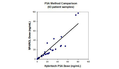
Figure 8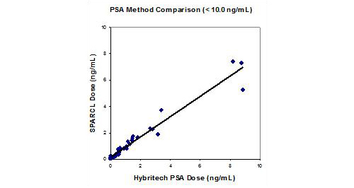
The examples of SPARCL solid phase formats exhibited both good sensitivity and good dynamic range (Figures 2, 3). The use of ascorbic acid to suppress background signal (Table 1) further enhanced sensitivity. When antibodies were directly labeled with acridan and used in a sandwich (Figures 4, 5, and 7) or a competitive format (Figure 6) a good sensitivity was achieved with a good dynamic range. Furthermore, it was demonstrated that a good correlation can be achieved with the reference PSA assay using solution phase reagents, especially at the clinically significant range.
SPARCL technology offers significant time savings and simplification of assay mechanics, which makes it an attractive technology for HTS and general life science research.
*Internal feasibility assays, not for commercial distribution.